Rabbit, Guinea Pig & Chinchilla Dental Care

Diet and Teeth – It doesn’t take much time around these animals to notice that their main activity during waking hours is eating. This is for very good reason; the health of their entire GI tract depends on this feeding style. They have adapted to specific nutritional sources and feeding habits over many generations. In general, the food they are adapted to eating is low in caloric density and high in fiber. The low caloric density means they need to eat a large volume to meet their caloric needs. The high fiber is important in stimulating normal coordinated movement of the intestinal tract. In captivity, it’s very important to have a good quality grass hay available at all times. Timothy, Orchard or a combination with some Oat Hay are all good choices. A handful of vegetables and a small amount of pellets helps ensure the full balance of vitamins and nutrients in the diet. Good dental health is a very important part of this process.
All of the teeth in these animals will continue to grow throughout their entire life. In addition to the incisors that are easily seen, they have between 16 and 22 teeth deeper inside the mouth that are more difficult to see. They all have 3 upper and 3 lower molars on each side. Guinea Pigs and Chinchillas also have 1 upper and 1 lower premolar on each side. Rabbits have 3 upper and 2 lower premolars on each side. These teeth must align properly to allow the muscles of the jaws to work effectively and to chew the food properly. If they are not properly aligned, the muscles may become painful or they may weaken. Additionally, malaligned teeth can develop sharp points along the tongue or cheeks that can cause pain during chewing. Teeth can also become abscessed, broken, or even develop masses. Unless these problems are properly identified and corrected, dietary problems will develop. This can be minor weight loss, or can be as serious as a life-threatening obstruction from improper chewing and poor muscle coordination in the GI tract.
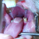
- Sharp tooth edges that can be appreciated along with a small cut on the side of the tongue was causing pain for this rabbit.
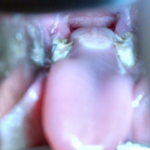
- Notice the sharp points on lower teeth against the tongue. After trimming with dental bit, rabbit was more comfortable.
A very important part of an annual (or semi-annual) examination by a veterinarian is a dental examination. At a minimum, physically palpating the jaws for abnormal shapes suggesting problems with the tooth roots will usually be followed by a look inside the mouth with the help of a cone or speculum and a light to look at the premolars and molars. As we will discuss further below, any abnormalities noted in the examination, or any suspicion of more serious concerns from the physical exam and history may need to have a sedated examination and some more advanced imaging to identify medical concerns.
Sedated exams and imaging
As you can imagine, looking at the teeth inside such a small mouth with an animal that chews (and holds) hay and doesn’t want a plastic cone in its mouth means that only a limited look is possible. That’s fine for just a routine screening at an annual health exam, but if there is indication of a concern, then a better examination is required. Sedation ensures that the mouth can be opened further and reduces movement that makes visualization difficult. It can also allow use of tools to spread the cheeks and tongue out of the way to get a better look at the sides of the teeth without risking harm to the pet. With the medications available in practice today, the reduced anxiety and stress with sedation can be far more safe for the pet than a less thorough exam without sedation.
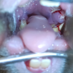
- A very sharp fractured tooth point which penetrated the cheek and caused an abscess. The fractured tooth was trimmed back, the sharp point removed and the abscess drained. Rabbit recovered well with antibiotics.
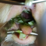
- The lower teeth of this guinea pig are angled inwards which can cause bridging that traps the tongue and prevents normal swallowing of food. After correcting the overgrowth, there can be rapid and drastic improvement.
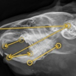
Sedation also allows imaging for what cannot be seen by just looking. 80-90% of the teeth in these animals live beneath the gums because they have very long toots. Any problem in the root can also translate into dental health problems and needs to be addressed. Radiographs allow a good look at the roots. They also allow measurements to be taken to help plan for correcting malalignment. By looking at the angles of the teeth and measuring relative to other structures in the head, specific plans can be formulated to trim overgrowth in a way that helps correct the alignment of the cheek teeth to correct the underlying problem.
In very complicated cases, 3-dimensional imaging may be recommended. Computed tomography (CT) scans can be done very quickly and will allow very detailed 3D images to be constructed. In cases that involve tooth roots erupting into other structures, cancerous masses, abnormal anatomy, large bone abscesses or complicated fractures, this can be very valuable in helping get a positive outcome. If a picture is worth 1000 words, then hundreds of pictures woven into a 3D image is priceless!
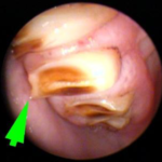
Detailed views of the teeth can be obtained using an endoscope. Many times even with sedation, problems in the back teeth can be difficult to see, as can be some problems on the sides of the teeth, or sockets where teeth are recently missing. Use of an endoscope can allow a magnified view of teeth and can allow some degree of looking around corners or seeing teeth and gums from different angles. Many times this tool has allowed a clear diagnosis and definitive treatment where other tools have failed.
Use of all of these tools appropriately allows the best opportunity to identify dental health problems and plan for effective treatment.
Treatment options for dental disease
Treatment, of course, depends on the condition diagnosed. The most common problem is overgrowth of the teeth. Again, radiographs should be done with rare exception. Before trimming the teeth, any contributing problems of the tooth roots need to be taken into consideration. Additionally, measurements and examination of the angles that the teeth oppose aid in planning which teeth to trim and how much they need to be trimmed. A dental bit is used to remove the excess tooth material just like drilling into a cavity. Unlike a cavity, only the enamel is removed. The pulp of the tooth is not affected, so there is not normally any pain from the procedure, though the muscles of the jaw may be sore when they are being used differently after the trim. If there are sharp points being removed, then there may still be some pain in the cheek and tongue from where the sharp teeth damaged them, but it will start to heal after the teeth are corrected.
- Example of measurements taken on radiographs to help plan the dental procedure. This is a guinea pig with overgrown lower molars and premolars.
- A view of the upper teeth through an endoscope. This allows very detailed images of individual teeth, even deep in the mouth.
Abscesses are another common problem. Generally, the infected area needs to be drained in order to allow antibiotics to effectively get into the area and deal with the infection. This can mean extracting a tooth, or if the deep root is affected, it may need to be drained from under the jaw. Samples of that material should be submitted to a lab whenever possible to identify the bacteria involved in the infection and target antibiotic treatment so that resistant infection can be avoided. Antibiotic medication can also be placed into the open socket after extracting the tooth by soaking it in absorbable material. This is especially effective if the endoscope is used to help visualize the socket.
Fractured teeth may be treated by trimming or extractions, depending on how bad the fracture is at the time of the procedure. Other less common conditions have a variety of possible treatments including surgical and medical procedures.
Healthy teeth are very important for healthy pets, and there are a variety of ways that problems can be properly identified, diagnosed and treated.
