Shoulder Instability & Pain in Dogs

Written by Staff Veterinarian
What is shoulder instability?
Shoulder problems are one of the most common causes of front leg lameness in dogs. Shoulder instability (SI) describes a wide range of soft tissue injuries to the ligaments, tendons and muscles of the shoulder. SI affects all sizes and breeds of dogs. Many athletic (esp. agility) dogs are affected, but more sedentary pets are also affected. At this time it is not clear if the condition is purely traumatic or a result of degeneration of the tissue. Typical patients are middle aged, but SI can affect all ages. SI most commonly affects a single leg, but a small percentage of pets have both shoulders affected.
How is SI diagnosed?
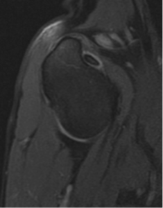
The starting point for diagnosis of SI is a thorough orthopedic and neurologic examination. Once pain has been localized to the shoulder, radiographs (x-rays) are typically the next step. It is important to note that radiographs typically do not diagnose shoulder instability, but are used to look for other causes of shoulder lameness (fractures, cancer, cartilage flaps or arthritis). Once radiographs are taken, the next step is generally a sedated measurement of shoulder stability (shoulder abduction angle) (Figure 1). Further diagnostic steps are dependent on this angle measurement. Often, ultrasound is used to evaluate the soft tissues of the shoulder.

Fig. 1: MRI demonstrating normal biceps.
In some cases, MRI (figure 2) may also be required to completely evaluate the extra-articular structures and determine the best treatment options. Cost may be rate limiting for these diagnostic tests. In most cases, exploratory arthroscopy must be performed to definitely diagnose the problem.
How do you treat SI?
Medical management: A variety of treatment options exist for SI and depend on the type and degree of instability present. For early cases, medical management may be the first step. Patients are sedated and fluid sampled from the affected joint. Long acting steroids (depomedrol 20-40mg) may be placed in the joint to decrease inflammation and pain.

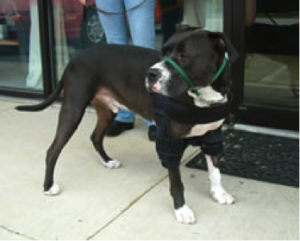
Fig. 2: Hobbles by Dogleggs.com
Patients will often be placed in a special type of hobble to limit shoulder motion during the healing phase (figure 3). Physical therapy is very important after the initial healing phase to regain shoulder strength. Treatments such as laser, therapeutic ultrasound and shockwave are often used during this phase of treatment.
Platelet rich plasma (PRP) is a new therapy that involves spinning down the patient’s own blood and harvesting the platelet portion of the plasma. This can be injected into the joint and may help stimulate the healing process. Objective evaluation of these therapies is lacking. Water therapy (swimming and water treadmill) can be very valuable with both surgically and medically managed SI in dogs.

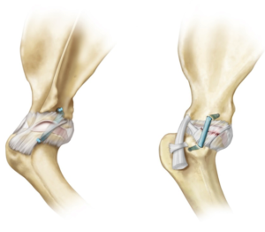
Fig. 3: Tightrope, minimally invasive shoulder reconstruction
Weight control is also very important. Overweight patients should be placed on appropriate calorie or metabolic diets. Medical management can result in good outcomes in about 2/3 of appropriately selected patients. Dogs with significant laxity (abduction angles >60 degrees) rarely resolve with medical management only. Surgical management: surgical management is the treatment of choice for more severe instability cases or in those that medical management has failed. A variety of surgical stabilization procedures are available and depend on the exact cause of SI. Minimally invasive surgical options are always considered. Ligament replacement surgery is often the surgical treatment of choice for SI (figure 4). Successful outcomes with surgery approach 90%. Patients can return to full function, but it may take 6-12 months to completely regain muscle strength. Complications with surgery are uncommon, but include infection, failure of the repair or persistent lameness.