Avian Respiratory System: Anatomy and Physiology
I hope that you find this fun to read. If it’s as fun for me to write and for you to read, then I plan to make this a periodic format for the website, so please send feedback to let me know if you are interested.
Let’s start the first episode with a doozy: the avian respiratory system.
Almost every aspect is unique compared to mammalian respiratory systems. As with most things I will publish in this series, there can be considerable species differences, but some generalities are very common. Like mammals, birds have nares, a larynx, trachea and lungs. In addition, they have nine air sacs and a syrinx (vocal center). Unlike mammals, they have no diaphragm and there is a unidirectional air flow that requires two full inspiratory and expiratory cycles to complete.

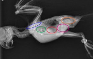
Relative locations of air sacs: Blue- cervical, green- clavicular (single sac medial), red- cranial thoracic, pink- caudal thoracic, brown- abdominal, grey- lungs.
Air enters through the nares, past the larynx and into the trachea (which has completely closed cartilaginous rings), past the syrinx at the bifurcation of the trachea into the bronchi. Down the bronchi, the air enters the caudal air sacs (2 paired caudal thoracic and 2 paired abdominal air sacs) with a small amount bypassing directly towards the lungs. At exhalation the air is expelled from the caudal air sacs into the ventrobronchi and dorsobronchi and down continuously narrowing airways in the lungs where gas exchange takes place. The exchange occurs in tubular air capillaries rather than saccular alveoli. The lungs are fixed anatomically and therefore do not expand or contract. At the next inhalation, the air moves into the cranial air sacs (paired cranial thoracic, paired cervical and a single clavicular air sac). At the next exhalation the air is expelled through the trachea once again. Since birds lack a diaphragm, the inhalation is achieved by expanding the chest, moving the ribs laterally, the sternum ventrally and cranially, and expansion of the abdominal muscles.
The syrinx differs from the mammalian vocal cords as well. There are no vocal folds. It’s located in the distal trachea and often into the bifurcation. There are vibrating membranes that are used to make sound. Variability between groups of birds is large, and beyond the scope of this article. Some of the bones of the wings and legs (species dependent) also have pneumatic centers connected to the air sacs. This helps dissipate heat during activities like flight that have a high metabolic demand. During flight the air sac expansion and contraction coincides with movement of the wings in many species for energy efficiency.
These anatomical differences make their respiratory systems incredibly efficient. There is continuous airflow across the air capillaries, so despite the relatively small size of the lungs, they are able to meet the incredibly high metabolic demands of birds in flight.
Clinical implications:
Capture and restraint can be stressful for any animal, and it seems to be one of the most intimidating things for new clinicians working with birds. I hear a lot of concern for killing birds by handling. Anatomy can help us with that. Closed tracheal rings allow for firm handling near the neck and behind the jaw without concern for choking relative to other animals. Firm handling around the head and neck means that there can be looser grip around the body. This is important since the ribs and abdominal muscles must expand to allow air sac expansion for respiration. Restraint with firm pressure near the head and loose pressure around the body helps with safety for the handler and the bird.
When evaluating respiratory effort, the expansion of the abdominal muscles and ribs can be a good indicator of labored breathing. Increased effort with the muscles compressing the caudal air sacs will result in a tail bobbing motion that is a good indicator of labored breathing. When a bird presents with tail bobbing or noticeable expansion of the ribs, it should be placed in oxygen and mild sedation should be considered.
The narrow air capillaries and efficient gas exchange means that birds are extremely sensitive to any airborne toxins (remember the canary in the coal mine). Owners should be cautioned about cleaning products, Teflon pans, perfumes, insecticides and anything causing new dust in the home such as construction work. Clinics should have a designated area for birds or cages that protect them from any cleaning products, fragrances used or any other airborne chemicals. Staff should be trained to think of birds especially closely when using any such products or dealing with strong odors.
Movement of air in the syrinx means it is a vulnerable area as well. This is a common place to find plaques from aspergillosis. The fungus likely gets easy hold in the area of the syrinx due to turbulent airflow and sensitive structure.
Air sacs can have stagnant regions that have vulnerability to infection. Significant infection can form around the lining of the air sacs with little radiographic change. There is also relatively little blood supply, making antimicrobial penetration difficult. Nebulization is often more effective for treatment of air sac infection. The anatomy does allow easy access for diagnosis and collection of culture samples with rigid endoscopy. Unrelated to respiratory disease, the size and location of the air sacs also allow good access to most major organs using rigid endoscopy. Direct visualization and biopsy sampling can be easily achieved with minimal invasiveness and a relatively short anesthesia time.
Finally, the compressibility of air sacs means there is vulnerability to restrictive airway disease. One of the most frequent causes of labored breathing is ascites causing compression of the air sacs from within the body cavity. Heart disease, reproductive disease, liver disease, neoplasia and numerous other conditions often present initially as labored breathing. Coelomic palpation should always be part of a standard triage exam for a dyspneic bird. Removal of fluid via centesis can provide rapid relief that may increase survivability. Removing even a small amount of fluid can relieve pressure against the air sacs and the vasculature, improving both ventilation and venous return.
Summary:
The unique anatomy and physiology of exotic animals is really fun to learn, but it also provides valuable information of clinical relevance. I hope this article has been interesting and informative even if your practice doesn’t include avian patients.