Patellar Luxation in Dogs
What is patellar luxation?
Patellar luxation (PL) is a common cause of hind limb lameness in dogs and also on rare occasions in cats. In this condition, the patella, or kneecap, is displaced from its normal resting place within a groove in the femur (thigh bone) and is instead located toward the inside of the leg (the medial surface) or outside of the leg (lateral surface). While this condition can occasionally be caused by traumatic injury, most affected animals are born with skeletal abnormalities that predispose them to developing this condition. Thus, this disease is typically diagnosed in young adult dogs. The degree of lameness seen is variable depending on the severity of luxation and underlying skeletal abnormalities.
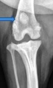
Figure 1A: Radiograph demonstrating the patella positioned centrally within the trochlear groove.
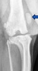
Figure 1B: Radiograph demonstrating the patella positioned outside of its normal position within the trochlear groove.
How is luxation of the patella (canine knee) diagnosed?
Typically the diagnosis of luxation (dislocation) of the patella is based on a thorough orthopedic evaluation. The limping seen in early stages is often intermittent, with the animal picking up the affected limb and walking on three legs when the patella is luxated and returning the limb to the ground when the patella returns to the groove. The severity of patella luxation is determined by an assessment of how easily the patella is luxated from the groove while the hind limb is extended or how challenging it is to return the luxated patella to its normal position within the groove. Radiographs (x-rays) can be taken to help confirm the abnormal position of the patella and to help rule out other causes of hind limb lameness (figures 1A and 1B).
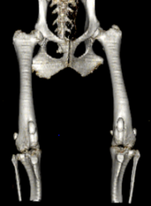
Figure 2: CT reconstruction of a dog with abnormal angulation and rotation of the tibia and femur, contributing to MPL.
In cases of advanced patella luxation, computed tomography (CT) evaluation with 3-D reconstruction is helpful in evaluating the limb for skeletal abnormalities that may contribute to luxation (figure 2). This reconstruction also aids in the surgical planning process.
Can other orthopedic diseases contribute to patellar luxation?
Patella alta, a condition where the patella sits too high within the trochlear groove, can be seen in association with patella luxation. The uppermost portion of the trochlear groove is shallower, allowing the patella to luxate more easily during normal rotation of the limb. Patella alta can be diagnosed on x-rays with the limb in a semi-flexed position. Correction of patella alta consists of a distal tibial tuberosity transposition, a surgical procedure during which the patella is moved downward.
How do you treat patellar luxation?
In early cases of patellar luxation where dislocation of the patella is infrequent or minimal lameness is seen, conservative management with rest and anti-inflammatory medications may be advised. In more advanced cases of MPL where the patella is difficult to return to its normal position or significant lameness is observed, surgical intervention is likely to be recommended. The board certified orthopedic surgeons at Ethos Veterinary Health hospitals perform several surgical techniques for repair of MPL in dogs including:
- Trochleoplasty
- Tibial Tuberosity Transposition
- Distal Femoral Osteotomy.
A combination of surgical techniques is often performed to achieve pain-free function of the operated limb.
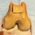
Figure 3: Trochlear block recession
Trochleoplasty
The goal of trochleoplasty is to widen and deepen the groove in which the patella normally rests. By creating a deeper groove that accommodates approximately 50% of the surface area of the patella, it is more challenging for the patella to luxate to the inside of the leg (figure 3). During this technique, the cartilage of the femur is preserved, and very little osteoarthritis is noted long-term.
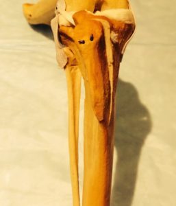
Figure 4: Tibial Tuberosity Transposition
Tibial Tuberosity Transposition
The patella is connected to the tibia along its tuberosity by a very strong ligament. The patella can be redirected to track within the groove by moving this ligament’s attachment on the tibial tuberosity toward the outside of the limb. This is accomplished by creating a controlled cut to the bone to allow the tuberosity to be placed in its new position. The tuberosity is then stabilized in the new position with wires or pins (figure 4). When correcting for patella alta, the tibial tuberosity is shifted downward (distally) to direct the patella to a deeper portion of the trochlear groove.
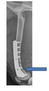
Figure 5: Radiographs performed immediately postoperatively of a DFO. Note the area where bone has been removed to allow the femur to be straightened (blue arrow).
Distal Femoral Osteotomy
In large-breed dogs diagnosed with patellar luxation, abnormal bone conformation can contribute significantly to luxation of the patella. These abnormalities lead to a bow-legged stance that increases tension on the patella, causing it to be drawn medially. The presence of these abnormalities is determined by evaluating radiographs, or a CT scan, to look for inappropriate angulation of the femur near the knee. To correct the angulation, a cut in the bone (osteotomy) is created to allow the limb to be straightened (Figure 5). A bone plate and screws are used to stabilize the osteotomy until healing is complete.
In general, the prognosis for return to pain-free function following surgery is good, with more advanced dislocated patellas having more difficulty in recovery. Following an initial 2-3 week period of extreme exercise restriction, a gradual increase in controlled activity is permitted; total recovery time following surgery is generally between 8-12 weeks. Your surgeon will review specific recommendations regarding postoperative rehabilitation during the initial consultation and follow-up visits. Postoperative physical rehabilitation, in some cases, can improve outcomes and shorten the recovery period.
Potential complications exist for any orthopedic surgery and include infection, implant failure, and incision complications. An experienced surgeon and appropriate postoperative care significantly decrease the risk and negative effects of these complications.