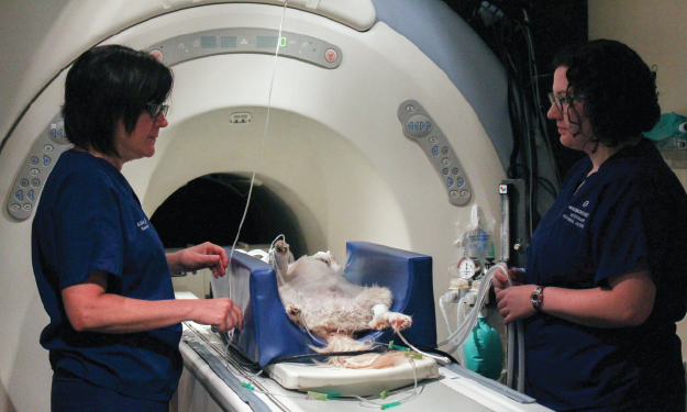
Magnetic Resonance Imaging (MRI) for Animals

MRI is the most common diagnostic imaging tool used in both human and veterinary medicine to visualize the nervous system. It is the current gold standard for imaging the nervous system in dogs and cats. It provides high quality images with excellent detail and resolution of the brain and spinal cord compared to other imaging tools, such as computed tomography (commonly referred to as a CAT scan).
Common examples of MRI use in animals include paralyzed dogs with disc hernaition, brain tumors, stroke, and many other conditions such as rupture cranial cruciate ligament.
MRI techniques for animals are exactly the same as those for humans. The imaging technique is exactly the same whether a “human MRI” machine or a “veterinary MRI” machine is used. The only difference between people and animals receiving an MRI is that animals will not lie still when asked. In general, it takes approximately 45 minutes to perform a MRI scan. Thus, animals must be under anesthesia for the scan. Prior to this, blood tests and X-rays often are performed to make sure that your pet is as safe an anesthetic candidate as possible.
MRI is very safe and complications associated with anesthesia are rare. The MRI is read by a board-certified neurologist for patients with brain or spinal cord disease or by our board-certified radiologist for non-nervous system MRI scans.