What Are The Signs of Hip Dysplasia In Dogs?
What is Hip Dysplasia?
Hip Dysplasia is a common, inherited, developmental condition that involves increased laxity of the hip joint. It is one of the most common orthopedic abnormalities in young, giant and large breeds, but all breeds can be afflicted. Although the exact cause of hip dysplasia has not been determined, many factors have been implicated. Genetics, rapid growth, excessive nutrition and diminished muscle mass have been associated with increased severity of hip dysplasia. Affected dogs are born with normal hips (Figure 1), but develop a lack of conformity between the femur and acetabular cup which invariably leads to the development of arthritis.
Figure 1: X-ray of Normal Hips
Diagnosis
Dogs with hip dysplasia may present with signs of hip pain, commonly indicated by a reluctance to jump into the car, pain when rising, or inactivity and reluctance to play as puppies. Signs are common between 6 and 18 months when there is excessive laxity (Figure 2) and again in dogs over 3 years of age when arthritis becomes more severe (Figure 3).
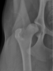
Figure 2: X-ray of Hip Dysplasia in Young Dog. There is increased laxity of the joint causing a separation of the femoral head (ball) from the acetabulum (socket) and there is already some arthritis forming along an area called the femoral neck.
Diagnosis is based on physical examination findings of laxity (a positive Ortolani test) or pain in the hips, and is confirmed with x-rays. We recommend screening examinations be performed by your regular veterinarian at four months of age in any large breed dog. Standard “hip extended” (OFA: Orthopedic Foundation of America) views combined with distraction views (PENN Hip) give the most information.
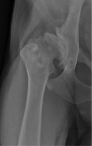
Figure 3: X-ray ofHip Dysplasia in Mature Dog. There is severe change to the bones making up the hip joint.
What are PENN Hip Radiographs?
PENN Hip has a strong scientific foundation as being the most effective hip screening tool for dogs. PENN hip is typically used on immature dogs to help determine their candidacy for breeding or help determine their risk of developing arthritis. The PENN Hip technique is performed on heavily sedated or anesthetized dogs and uses a padded distraction device placed between the back legs while the x-ray is taken. Evaluation of these images will generate a distraction index (DI) that will help give an objective assesment of the degree of hip laxity present and help predict the likelyhood that your pet will develop arthritis in the future. Dogs with a distraction index of less than 0.3 are very unlikely to develop arthritis, while those with a DI greater than 0.7 are highly likely to develop arthritis. PENN hip can be performed as young as 16 weeks of age. It has been shown that breeding dogs with tight hips (low DI) to dogs with tight hips can decrease the incidence of hip dysplasia.
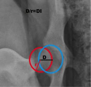
Figure 4: PENN hip radiographs demonstrating how DI is determined.
Treatment
Many treatment options are available for hip dysplasia. Treatment is based on severity of signs, age at presentation, your expectations of performance, and financial considerations.
Medical Management
The most conservative method of treatment of hip dysplasia involves medical management consisting of weight loss, controlled activity, physical therapy and anti-inflammatory drugs or nutraceuticals (e.g. glucosamine, omega 3 supplemenation, see our link on osteoarthritis). Many new non-steroidal anti-inflammatory drugs are available for dogs. Ask your family veterinarian which are best suited for your pet’s needs. Medical management does not reverse arthritis, but provides control of pain. Weight loss can be as effective as medications at decreasing pain and improving quality of life.
Symphysiodesis
Symphysiodesis is a technique for preventative management of the progression of juvenile canine hip dysplasia. It involves closing the growth plate on the “floor” of the pelvis, increasing femoral head coverage as patients grow. This technique can be performed relatively rapidly, is not highly invasive, and entails no surgical implants. Ideal candidates have hip laxity with no radiographic signs of arthritis and are between 15 and 20 weeks of age. Complications with the procedure are rare but include infection and incision complications. Most patients can be discharged the day of surgery and recovery is complete by 2 weeks after the procedure.
Triple Pelvic Osteotomy (TPO):
Dogs that have hip laxity but not significant arthritis are candidates for Triple Pelvic Osteotomy (TPO). Because arthritis can progress fairly quickly, dogs that are candidates for TPO are typically between 6 and 12 months old. TPO improves femoral head coverage (Figure 4) through a procedure that involves making three cuts in the bones of the pelvis, then rotating and plating a section of the pelvis. The goal of this surgery is to decrease pain and the progression of arthritis. Long term evaluation of dogs after TPO has shown excellent results. Complications include infection, anesthetic risk, incision complications and implant complications. Complications after surgery are uncommon with the new locking, titanium plate system that we utilize.
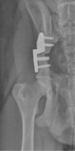
Figure 5: X-ray of TPO with Securos locking plate
Femoral Head and Neck Ostecotmy (FHO)
FHO is a salvage procedure to address hip dysplasia. This surgery involves removing the bone of the femoral head (Figure 5) and aims to eliminate the source of pain. Small dogs and cats may be better candidates for this procedure than large and giant breed dogs which may have prolonged recoveries and variable outcomes. Gait abnormalities may persist after FHO and post operative physical therapy is critical to the success of the procedure.
Total Hip Replacement (THR)
THR involves implantation of a prosthetic hip in a similar fashion as is done in humans (Figure 6) and has been well – established in veterinary medicine since the 1970s. Both “cemented” and “cementless” options are available for dogs and your surgeon will discuss which would be best for your pet. The availability of many different implant sizes makes this the best option for most dogs with severe signs of hip problems. Ideal candidates are over six months of age with no overt systemic illness. Although hip dysplasia is often a bilateral disease, THR is performed on only one hip at a time, with the most painful hip treated first. Often, only one hip needs to be replaced to achieve acceptable function. If pain persists, a second THR may be performed at least two months after the first. As with any surgery, complications exist with THR and include infection, dislocation, implant failure and femur fractures. These complications combined occur in less than 10% of patients and improvements in implant design and technique have led to the low complication rate. Full recovery from THR takes approximately eight weeks with most dogs able to walk on the affected leg the day after surgery. The success rate of THR is excellent with 90-95% of dogs able to have normal use of the affected limb after surgery.
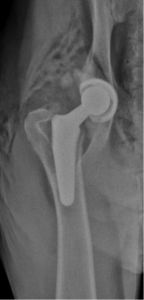
Figure 6: Cementless hip replacement
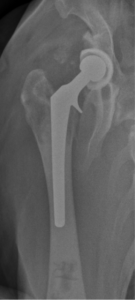
Figure 7 Hybrid (cemented femur, cementless acetabulum)