Lump on Dogs Elbow: Cancer Diagnosis & Treatment

Written by Marielle Goossens, DVM, DACVIM, Noelle Bergman, DVM, MS, DACVIM (Oncology), and Kendra E.F. Knapik, DVM, DACVIM (Oncology)
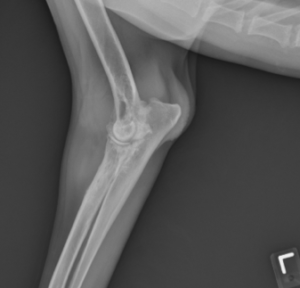
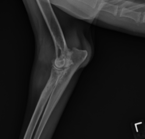
Phineas, a 7-year-old Flat Coated Retriever, was presented to Peak for evaluation of left thoracic limb lameness of 10 month duration. During physical examination, marked soft tissue swelling and decreased range of motion of the left elbow was noted. Radiographs of the left elbow showed marked bone proliferation associated with the elbow joint. The radiographic and physical exam findings were most consistent with a mass arising from the elbow joint. Since neoplasia was the top differential diagnosis for a mass in the elbow of a middle-aged Flat Coated Retriever, thoracic radiographs were ordered. No evidence of intrathoracic metastasis was observed. A CT scan was performed next, and a mass enveloping the left elbow was confirmed. An incisional biopsy of the mass was taken prior to recovering Phineas from anesthesia.


The histopathologic diagnosis was a sarcoma, and the pathologist was most suspicious that this tumor was of histiocytic origin given the signalment of the patient, tumor location, and the microscopic features of the biopsy. The mitotic index was 14, and there was marked anisocytosis and anisokaryosis with multifocal cytomegaly, karyomegaly, and multinucleated giant cells. The neoplastic cells extensively invaded the adjacent skeletal muscle. Other differentials for a tumor arising from the joint that were considered included a synovial cell sarcoma and other soft tissue sarcomas. Phineas was referred to the Oncology Service for consultation regarding the biopsy findings.
Diagnostic Work Up
Special stains were ordered to confirm the diagnosis of histiocytic sarcoma. The neoplastic cells were diffusely positive for both CD18 and CD204, which are two markers for cells of histiocytic origin.
Histiocytic sarcoma (HS) is a malignant neoplasm of histiocytic cells. Dog breeds that are overrepresented with this neoplasm include Bernese Mountain Dogs, Flat-Coated Retrievers, and Rottweilers.These tumors can present in two main forms: localized and disseminated. The localized form involves a single primary tumor arising from a joint, cutaneous or subcutaneous tissue, lung, or essentially anywhere in the body. Patients with the disseminated form present with an advanced stage of disease and often with multiple visceral organs involved. Most commonly the liver, spleen, lungs, and lymph node are affected. The prognosis for dogs with the localized form of the disease is significantly better than the disseminated form. The localized form of HS has been shown to have a median survival time beyond 500 days following treatment compared to several months with the disseminated form. Commonly, patients with the disseminated form present with signs of systemic illness including weight loss, anorexia, vomiting, and occasionally, fever. Despite the superior prognosis, localized histiocytic sarcomas still have a high risk of metastasis to lymph nodes, lung, liver, spleen, and other organs.
Treatment and Follow-up
Given the high risk of metastasis, an abdominal ultrasound was recommended for Phineas. No evidence of abdominal metastasis was observed. Since Phineas’ disease was confined to his elbow based on the staging tests, a left forelimb amputation was recommended followed by chemotherapy with CCNU (lomustine). Periarticular forms of HS have been shown to have a significantly improved prognosis over HS arising from other organs. Periarticular HS that has not metastasized has been shown to have a median survival time of 980 days following treatment in one study. While amputation is the treatment of choice, palliative radiation therapy can also be used to help control pain in patients that are not candidates for amputation.
CCNU is the chemotherapy drug of choice for HS. The response rate in the gross disease setting has been reported to be 40-60%. For patients that develop progression of their disease with CCNU, doxorubicin with zoledronate (a bisphosphonate) is another option. This combination of drugs has displayed synergistic cell death in in vitro studies evaluating HS. Anecdotally, impressive responses have been observed with this drug combination in practice as well.
Phineas recovered well from his forelimb amputation despite concurrent orthopedic diseases (elbow dysplasia and history of a TPLO on a hind limb). His orthopedic diseases were managed with a combination of carprofen, gabapentin, amantadine, and Dasaquin. Following limb amputation, Phineas was started on chemotherapy. He received 6 doses, which were administered every 3 weeks. Restaging tests with thoracic radiographs and abdominal ultrasound were performed in the middle of the chemotherapy protocol and again at the end. No evidence of metastatic disease was observed at either time point. We have continued to monitor Phineas with these staging tests every 3 months. Phineas was diagnosed in August 2016 and was clear of disease on his most recent staging tests.
REFERENCES:
- Withrow, S.J., D.M. Vail, R.L. Page. Small Animal Clinical Oncology. St. Louis: Elsevier Saunders, 2013. Print.
- Klahn, SL, BE Kitchell, NG Dervisis. Evaluation and comparison of outcomes in dogs with periarticular and nonperiarticular histiocytic sarcoma. J Am Vet Med Assoc 239.1 (2011): 90-96.
- Skorupski, KA, et al. CCNU for the treatment of dogs with histiocytic sarcoma. J Vet Intern Med 21.1 (2007): 121-126.
- Skorupski, KA, et al. Long-term survival in dogs with localized histiocytic sarcoma treated with CCNU as an adjuvant to local therapy. Vet Comp Oncol 7.2 (2009): 139-144.
- Hafeman, SD, D Varland, SW Dow. Bisphosphonates significantly increase the activity of doxorubicin or vincristine against canine malignant histiocytosis cells. Vet Comp Oncol 10.1 (2012): 44-56.