Corneal Ulcers in Dogs

Written by Nancy Thompson, CVT
Eye issues are taken very seriously in veterinary medicine. When we hear that a pet’s eye is squinting, red, swollen, irritated, appears glossy, or is producing discharge, we encourage pet owners to go directly to a veterinary hospital to have it assessed. Sometimes, it is just a simple eye infection which can be treated with antibiotics.
But sometimes, it can be a more serious underlying disease or injury such as glaucoma, cataracts, or corneal ulcers. These more advanced eye issues progress quickly and if gone untreated, they can cause serious damage or blindness. Corneal ulcers are particularly common in small animal medicine and depending on how deep it is, can be very dangerous for your pet.
The Cornea: The Window of the Eye
The cornea is the transparent window that makes up the front surface of the eye. It is composed of three main layers:
- Epithelium– a thin layer of cells on the outer surface of the cornea
- Stroma– the main supportive tissue of the cornea and makes up 90% of its thickness
- Descemet’s membrane– the deepest and very thin layer; on the other side of Descemet’s membrane is the aqueous humor, the clear fluid that fills the inner chamber of the eye
Corneal Ulcers: Sometimes Serious, Sometimes Not
A corneal ulcer is a wound or abrasion on the corneal surface. A superficial corneal ulcer involves only the surface epithelium. This is a much less serious injury, but still requires veterinary care.
A deep corneal ulcer begins to involve the corneal stroma. The presence of a deep corneal ulcer usually indicates that a bacterial infection is present. The bacteria release substances that degrade the corneal stroma, causing the ulcer to progress deeper. If the ulcer extends to the deepest level of Descemet’s membrane, this is referred to as a descemetocele and is considered a serious emergency due to risk of rupture of the eye. If Descemet’s membrane ruptures, the fluid inside the eye leaks out and can potentially lead to irreparable blinding damage to the eye.
Causes: Most Commonly Trauma
There are several causes for corneal ulcers in dogs, and they occur most commonly due to trauma to the eye. Superficial corneal abrasions can occur from physical or chemical trauma such as:
- Rough play with other dogs
- Running through heavy vegetation or woods
- Irritating substances such as shampoo or dust/debris
- Infection or bacteria (less common)
More serious injuries to the cornea can occur from lacerations including:
- A cat scratch or a sharp object
Corneal ulcers are also common in certain breeds or dogs with underlying diseases such as:
- Dry eye, where decreased production of tears leads to drying of the corneal surface
- Brachycephalic (flat-faced) breeds with prominent eyes (such as Boston Terriers, Pugs, Shih Tzus, Boxers, and Bulldogs)
Symptoms: It is Painful
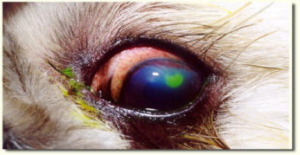
Eye stain performed on a dog
Corneal ulcers symptoms are painful and you may notice that your dog is squinting, pawing, or rubbing at the eye. Other symptoms can include redness and excessive discharge or tearing. Superficial ulcers aren’t typically visible to the naked eye, and your veterinarian will look for the presence of an ulcer with a special stain called fluorescein. When the stain is placed on the eye, the dye will adhere to the ulcer and produce a green fluorescence that identifies the presence of the ulcer.
Treatment: Medication vs. Surgery
Simple superficial corneal ulcers will heal on their own without incident in 3-10 days depending on the size of the ulcer. While the healing process takes place, treatment for simple superficial corneal ulcers includes antibiotic eye drops or ointments to prevent infection, as well as pain medications to relieve discomfort. Your pet will need to wear an E-collar (cone) to protect the eye during the healing process, as self-trauma to the eye can delay and complicate healing.
In the case of a deep corneal ulcer, more aggressive treatment will be necessary to prevent the ulcer depth from progressing. This typically includes multiple eye drops given several times a day, as well as pain and anti-inflammatory medications by mouth. In severe cases, referral to a veterinary ophthalmologist for corneal surgery to place a graft onto the ulcer may be necessary to stabilize the cornea and prevent rupture of the eye.
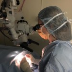
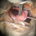
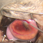
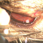
a keratectomy on a patient after a corneal ulcer worsened and removal of the diseased layers of the cornea was needed:
Healing: Follow-up Care is Crucial
Follow up with your veterinarian is needed to determine if your pet’s ulcer has completely healed. You should continue treating your pet with all prescribed medications until your veterinarian indicates that the ulcer is fully healed. Simple superficial corneal ulcers should heal within 1-2 weeks or less, however in some cases the ulcer may be slower to heal.
If your dog’s ulcer doesn’t heal or show signs of healing within this time frame, this indicates that an underlying cause may be present (dry eye, abnormally-directed eyelashes, entropion, etc.) or that additional procedures may be needed to facilitate healing of the ulcer. In older dogs, chronic non-healing (indolent) corneal ulcers are very common and often require additional procedures such as a diamond burr debridement (see image below) to encourage and facilitate healing. If your pet’s corneal ulcer is not healing appropriately and an inciting cause cannot be identified, your veterinarian may recommend referral to a veterinary ophthalmologist for specialty care.