Cataracts in Poodles

The Poodle
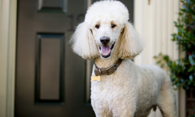
The Poodle as a breed has its origins in Germany and likely dates back to the 15th and 16th centuries. The larger Standard Poodle was widely used as a hunting dog, and was known especially for being a strong swimmer. The classic haircut we often see on poodles was actually invented to improve dog’s performance in the water. The smaller poodles came along only shortly after the standard poodle, but were not used for hunting. This smaller version found its way onto the laps of owners, or was used for finding truffles in the woods.
Cataracts – Not just a problem for Poodles!
The vision limiting or blinding process of cataract development is a concern in the Poodle breed (mostly the miniatures), but also affects many other breeds such as Cocker Spaniels, Labrador and Golden Retrievers, Boston Terriers and Bichon Frise, to name a few. To understand the cataract condition, it helps to know something about it when it is normal. Structurally the lens is just like a plain M&M that sits behind your iris. It has an outer shell and an inner filling. It’s a ridiculously simple concept, but an apt description. The outer shell and inner filling are clear when all is well. When a cataract forms, most commonly, the filling becomes white. Since the lens inside the eye is responsible for transmitting and focusing an image onto the retina, when it loses that clarity it fails to function properly and the animal or person loses vision.
If your pet is losing vision you may notice him or her being less active, moving around more cautiously, not playing as well, or in the advanced state, becoming completely blind. Cataracts are one potential cause of this vision loss. They are diagnosed by dilating your pet’s eyes, just like you would have done for your own eye examination. The clarity of the lens and very importantly the health of the retina are examined with a slit lamp biomicroscope and ophthalmoscopy. At times ocular ultrasound and electroretinography are necessary. All of this medical equipment is standard among veterinary ophthalmologists. The full examination is necessary to ensure the cataract is the cause of vision loss and that the retina is normal. I like to say that there is no reason to perform cataract surgery if the retina isn’t healthy. Remember: your lens transmits and focuses images, your retina makes sense of it all.
Correcting Cataracts
To correct this cataract condition, surgery is necessary. Cataract surgery is nearly identical to the surgery performed for people. Going back to the M&M example, the surgery effectively removes the white filling and leaves the clear outer shell intact and in place inside the eye. Once the filling of the lens has been removed you lose your ability to focus on near objects. So to bring your world into better view, a new, clear, artificial lens is inserted into the empty shell/capsule of the original lens. Now both transmission and focusing of light can occur, and the patient/dog/pet can function normally again. It’s a simple concept. In practice though this is detailed microsurgery that is performed with the assistance of general anesthesia. The outer shell of our lens is fractions of a millimeter thin. The whole surgery is performed inside the eye and through the pupil. Manipulating tissues like this is ultimately fine surgical work and can only be performed by a veterinary ophthalmologist.
So is it even worth it to pursue. I strongly believe, yes. Reported success rates of 95% are most common. Animals come in unable to be the pets we want because of vision loss and they can return to full function. This may be spotting decoys during field trials, chasing the ball on the beach, or zipping confidently through the woods. It is a privilege to be able perform this surgery for our companions. The boost in quality of life is amazing.