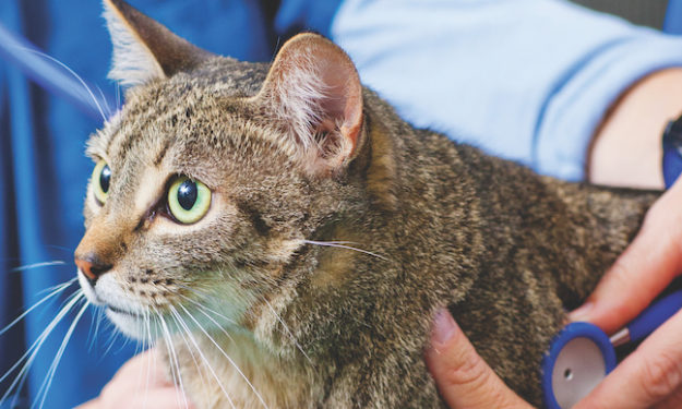
Case Study: Feline Chronic Renal Disease vs Hyperaldosteronism

History
Melvin, a 9 year old male neutered DSH, presented to the Veterinary Specialty Hospital for evaluation of an acute onset of ataxia and cervical ventroflexion. He is strictly indoors with no history of toxin exposure, no travel history, and no significant prior medical history.
Physical Exam Findings
On presentation, Melvin was bright, alert and hydrated with mild cervical ventroflexion. His signs quickly progressed to marked cervical ventroflexion, generalized weakness, reluctance to walk, and a plantigrade stance with prolonged standing. The remainder of his neurologic examination was unremarkable.
Differential Diagnoses
Melvin’s neurologic signs are consistent with neuromuscular disease. Differentials for generalized neuromuscular disease include hypokalemic myopathy, myasthenia gravis, infectious or inflammatory myopathy, toxin, and less likely a vascular event. Differentials for hypokalemia include hyperaldosteronism, chronic renal disease, metabolic alkalosis, dietary deficiency, concurrent insulin therapy, and use of medications such as penicillins, amphotericin B, and loop diuretics.
Diagnostics
A complete blood count was unremarkable. A chemistry panel showed hypokalemia (3.2 mmol/L), mild azotemia (BUN 59 mg/dL and creatinine 2.9 mg/dL), hypercalcemia (11.1 mg/dL), and hypophosphatemia (1.7 mg/dL). A urinalysis showed a USG of 1.026 with 4+ blood, few epithelial cells, and few calcium oxalate crystals. The urine potassium concentration was elevated (69.7 mmol/L). A systolic blood pressure was mildly elevated (170 mmHg), however subsequent measurements were normal. Abdominal ultrasound exam revealed moderate bilateral chronic renal changes and multifocal hyperechoic splenic nodules most consistent with benign myelolipomas.
Due to the possibility for hyperaldosteronism as a cause for Melvin’s hypokalemia, an aldosterone level was submitted and was elevated at 1,043 pmol/L (reference range 194-388 pmol/L).
Plan
Melvin was treated with intravenous fluids containing potassium chloride and potassium phosphate supplementation. Overnight, he remained stable, but his neurologic status did not improve. Despite fluid therapy, his potassium level decreased to 2.3 mmol/L. His intravenous potassium supplementation was increased and oral potassium gluconate was started. His phosphorous level increased to 8.6 mg/dL, and potassium phosphate supplementation was subsequently discontinued. Serial blood work showed marked improvement in acid-base balance and potassium levels, with normalization of his potassium level and renal values within 72 hours. His intravenous fluids with potassium chloride supplementation were tapered and he continued to do well. His cervical ventroflexion resolved and his overall strength improved. He was then discharged with instructions to continue oral potassium gluconate BID and switch to a renal diet. He was rechecked three days later, and was clinically doing well at that time. Recheck blood work showed normokalemia (3.95 mmol/L). He was rechecked two weeks later, and was continuing to do well. Recheck blood work showed normokalemia (4.2 mmol/L), mildly elevated BUN (51 mg/dL), and normal creatinine (1.6 mg/dL). A repeat aldosterone concentration was below the reference range at 67 pmol/L.
Discussion:
Hypokalemia is typically defined as a serum potassium concentration below 3.6 mmol/L. Treatment depends on the severity of hypokalemia and associated clinical signs including polyuria, polydipsia, ileus, and muscular weakness. Potassium can be supplemented both intravenously and orally, and frequent measurement of potassium concentration is essential to assess response to therapy and avoid iatrogenic hyperkalemia. The primary cause for the hypokalemia should also be identified and treated appropriately.
Hyperaldosteronism generally reflects the appropriate physiologic response to counteract hyponatremia, hyperkalemia, and hypotension. In Melvin’s case, we did not know initially if the hyperaldosteronism was secondary to an aldosterone-secreting adenocortical adrenal tumor (Conn’s syndrome) or secondary to renal disease given his azotemia. There was no evidence of an adrenal tumor noted on his abdominal ultrasound exam, although changes are not always apparent. Cats with adrenal tumors will have aldosterone concentrations ranging from 800-14,000 pmol/L and most have concentrations greater than 1000 pmol/L. Cats with renal disease can have aldosterone concentrations ranging from 130-1,670 pmol/L. These cats will typically have bilateral micronodular hyperplasia of the zona glomerulosa of the adrenal cortex and have ultrasonographically normal adrenal glands.
Due to the overlap of aldosterone concentration ranges between these two diseases, interpretation of concentrations can be challenging and other clinical parameters must also be evaluated, including electrolytes, blood pressure, and ultrasound examination of the adrenal glands. In Melvin’s case, we rechecked an aldosterone level approximately two weeks later and it was below the reference range.
This finding in combination with his normal adrenal ultrasound, normal blood pressure, and improved azotemia would suggest chronic renal disease as the cause of his previously documented hyperaldosteronism.
Melvin is continuing to do very well clinically and loves his new renal diet. We are gradually tapering his potassium gluconate supplementation and his renal values remain stable.