Broken Bones in Pets: Fracture Repairs for Dogs and Cats
If you are reading this, it is likely you received the devastating news that your pet is suffering from some unfortunate trauma. We know this can be an incredibly stressful and traumatic time for both you and your pet. How will things turn out? Will he/she be OK?
There are many factors that will affect the answer to this question. The severity and nature of the injury is certainly one of them. Equally important is the skill, experience, and tools of the team treating him/her. The success of your pet’s fracture treatment has much to do with type and severity of the injury, but also with the experience and equipment of the team treating them.
No two fracture cases are exactly the same, but it is unlikely that the teams of surgeons at our hospitals have not seen many fractures similar to the one you are facing.
What to do if your pet breaks a bone
After an accident, it is important to ensure your pet is stable and not experiencing major internal injury. You will likely start at an ER or at your pet’s primary care veterinarian, and end up consulting with a surgical specialist.
A discussion with the surgeon will determine the need for testing like blood work, x-rays of the chest, or an ultrasound of the abdomen to look for additional trauma. Once it is time to focus on fracture repair, we will take a lot of different factors into consideration when determining the best repair technique for your pet.
How are fractures treated?
A variety of treatment options exist for repair of fractures. This depends on the type and location of fracture, the age and activity of the patient, the presence of other injuries and your ability to manage your pet in the post-surgical period. There are often many routes to healing for each case, and the goal is to pick the intervention that will result most consistently in the fastest healing with the least amount of complications in the short and long term.
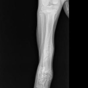
This is approximately 3 weeks from the injury. The bone is healed enough to allow removal of the splint. The bone will continue to heal and “remodel” to a more normal appearance.
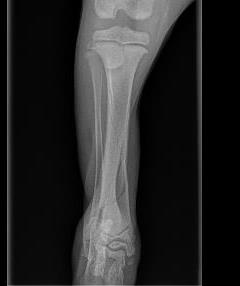
3 month old Great Dane puppy with a tibial fracture. This is a cranial-caudal or “front-to-back” view showing the fracture at the lower (distal) portion of the tibia (shin bone). Note that the small fibula bone that runs parallel to the tibia is not fractured. In a young patient, this fracture can heal successfully with an appropriately-placed and maintained splint or cast.
Splints and casts for pets
Also known as external coaptation, this form of treatment is typically used for fractures that are closed (not poking through the skin), below the elbow or knee, not significantly displaced, and usually not more than two pieces. A variety of fractures can be managed successfully with splints, but it is important to remember animals have a hard time telling us when there is a problem with their splint.
It is also very hard to convince our animal patients to use crutches and stay off of their splinted limb while it heals. Bandages that get wet, slip, are too loose or too tight, can all cause devastating complications. When factoring in the decreased initial cost of doing a splint owners should be aware of the typical weekly bandage changes, possibly with sedation, and the need for follow up x-rays.
Bone Plates (xray of bone plate radius, femur, humerus, plate rod, picture of lockings)
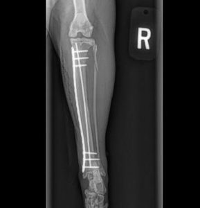
Radiograph immediately after surgery showing a “plate-rod” repair, restoring the alignment of the bone and providing stability to allow bone healing without the need of a cast or splint.
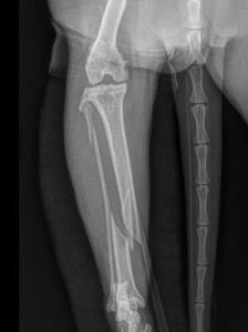
Fractured tibia in a cat.
Bone plates and screws
One of the most common methods of fracture repair is the application of a metal bone plate and screws to bridge the fracture. This is an extremely versatile method of repair and can be used in everything from the smallest puppies and kittens to the largest giant breed dogs. The benefit of plates and screws is that they allow early use of the operated area, particularly the joints around the injury, and often avoid bandages with their associated complications. Early limb use helps prevent muscles from atrophying, keeps the joints healthy, and helps to stimulate bone healing.
It is important to realize that patients will often feel back to normal within a couple of weeks from surgery, but it will take the bone 6-12 weeks to heal. During that period, controlled activity is extremely important to prevent breakage of the implants.
Complications after fracture repair include bending or breakage of the plate and screws, infection, swelling and incision complications. We generally leave bone plates on for the rest of the patient’s life even though they are no longer needed once the bone has healed. Patients that develop an infection may need the bone plate to be removed after the bone is healed to resolve the infection.
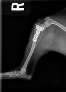
Radiograph immediately after surgery. The bone has been returned to its normal position with a specially-designed titanium locking plate that acts as an internal fixator on the bone.
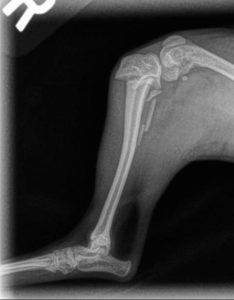
Fracture of the upper portion of the tibia in a Chihuahua. This injury would have a guarded prognosis with a cast or splint.
External Fixators
External fixators are a common method of fracture repair. Metal pins are placed through the skin and into the bone. The pins on either side of the fracture are connected to a variety of different connecting bars made up of acrylic, carbon fiber, stainless steel or titanium. External fixators are an extremely versatile method of repair and can be used on everything from fractures in the mouth to fractures of the spine.
External fixators can often be placed with a minimally invasive technique without having to open the fracture site and disrupt the initial healing that has already started from the moment of the injury. We often do this type of repair for very young patients with fractures of the tibia or for open (bone poking through the skin) fractures, because plates placed in open fractures may be at higher risk of harboring infection. We will often use fluoroscopy (moving x-ray) to assist us in placing external fixators to minimize the need for surgical exposure.
An additional benefit of external fixators is that they can be easily removed once the bone has healed without the need for an additional surgery. The drawback is that there is more follow-up (weekly) and there is an apparatus attached to the patient. Often patients will need to wear an e-collar until the apparatus is removed. Complications are similar to plates such as infection, implant failure, and failure to heal of the fracture.
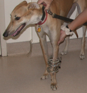
Ring fixator for fractures close to joints or limb deformity.
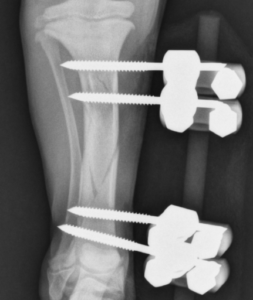
External fixator in a young patient.
There are times when an internal implant like a plate, pin, screws, or combination of those, can be placed non-invasively as described above. In the right situation, this can provide many advantages of both non-invasive surgery (smaller incisions, less trauma, precise implant placement using fluoroscopy) and avoid the issues with an external apparatus. Minimally-invasive internal fracture repair can be used for fractures of the long bones (like tibia), certain elbow fractures, and separation of the pelvis from the spine (sacroiliac fracture/luxation). This is another example of VSH’s cutting-edge approach to fracture repair.