Elbow Dysplasia in Dogs
Osteoarthritis of the elbow joint is the most common cause of foreleg lameness in dogs. Most of the arthritic diseases of the elbow are considered forms of developmental elbow malformation (dysplasia).
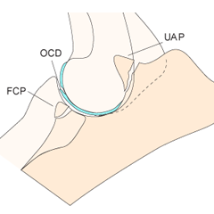
Elbow dysplasia refers to a group of congenital diseases of the elbows of dogs, which include:
- Fragmented coronoid process (FCP)
- Medial compartment disease (MCD)
- Osteochondrosis dessicans (OCD)
- Ununited anconeal process (UAP)
- Incomplete ossification of the humeral condyle
FCP and Medial Compartment Disease
Fragmented coronoid process (FCP) is the most common form of elbow dysplasia in dogs. In this disease, a fragment of bone and cartilage of one of the bones of the elbow joint (ulna) is broken off. The rest of the joint may be normal, or there may be additional cartilage damage, including OCD or severe full-thickness cartilage loss. This is termed Medial Compartment Disease and, unfortunately, can occur in dogs as young as 1 year of age.
FCP and medial compartment disease are best diagnosed and initially treated with arthroscopy. Arthroscopy is the fastest, most effective, and least invasive method for fragment removal. Advanced medical options for treatment of Medial Compartment Disease include joint injections with Hyaluronan or autogenous Stem Cells.
Advanced surgical treatments of Medial Compartment Disease include Sliding Humeral Osteotomy (SHO) and total elbow replacement. Dr. Schulz developed Sliding Humeral Osteotomy in the Orthopedic Research Laboratory of the University of California and is currently in clinical trials with very positive results.
Osteochondrosis Desiccans
Osteochondrosis dessicans (OCD) is an abnormality in the development of cartilage that leads to a cartilage flap. In the elbow this occurs on the humerus and can usually be detected on radiographs.
Treatment of elbow OCD involves removing the loose cartilage flap by arthroscopy. Removal of the cartilage flap may enable the underlying bone to heal with fibrous cartilage tissue, stopping the irritation of the opposing cartilage surface. OCD is best treated by elbow arthroscopy.
Ununited Anconeal Process
The anconeal process is the top part of one of the bones of the elbow called the ulna. In some dog breeds, especially German shepherds, this fragment of bone may fail to unite with the rest of the ulna during a puppy’s growth in the first year of life. When this occurs, the loose fragment contributes to joint instability and inflammation.
Diagnosis of ununited anconeal process is easily made with x-rays in dogs older than 6 months. Treatment involves surgical techniques to either remove or stabilize the bone fragment. The key to successful surgery of ununited anconeal process is early diagnosis when the osteoarthritis is not yet severe, and the body is still able to heal the fragment to the remainder of the bone.
Incomplete Ossification of the Humeral Condyle
Incomplete Ossification of the Humeral Condyle (IOHC) is an uncommon disease of the elbow joint seen most often in Spaniel breeds. In this disease two of the parts of the humeral bone fail to unite. The end result is a permanent crack in the upper bone of the elbow joint. IOHC is very difficult to diagnose on x-rays because the crack in the bone is very narrow. The crack is, however easily seen by arthroscopy. Treatment of IOHC is to place a screw across the bottom of the humerus (humeral condyle) and the bone crack to stabilize the bone and prevent a future fracture.
Minimally Invasive Surgery
Arthroscopy is performed by a highly experienced and caring surgical team who prioritize your pet’s welfare. Our philosophy is to educate owners and help them make the best decisions for their pet and their family.
Arthroscopy is an advanced surgical technique for examination and treatment of joint disease. In arthroscopy the surgeon makes a small incision and inserts a pen sized instrument (arthroscope) to work inside the joint. Arthroscopy provides better visualization and greater opportunities for treatment of joint disease while being much less invasive than traditional surgery. Recovery from surgery is quicker, the incision is smaller and in most cases patients will not need to spend the night at the hospital.